- CE MDR
Dermus · Hungary
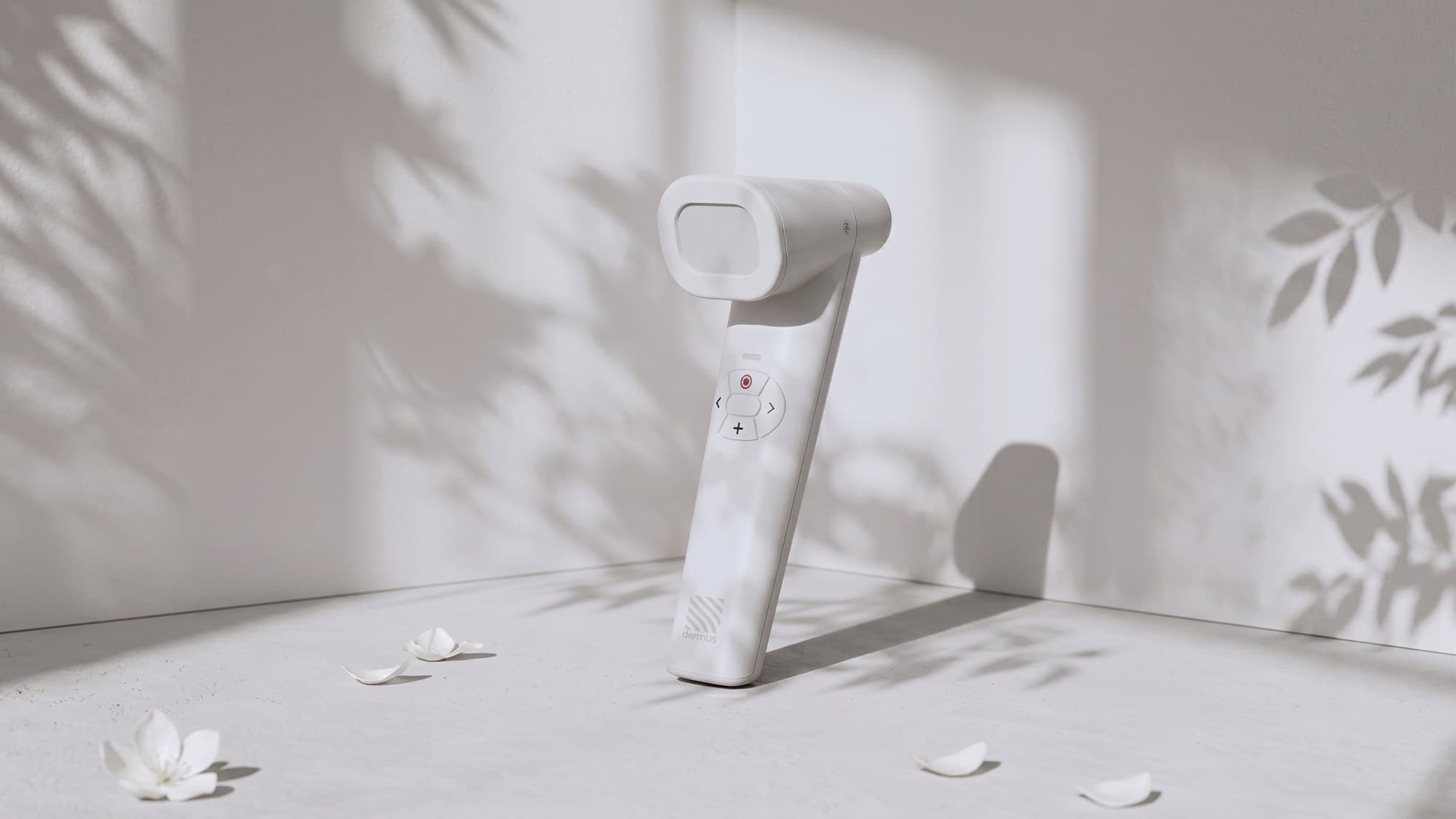
Dermus skin scanner
Optically guided high-frequency skin ultrasound
- Dermoscopy-guided ultrasound, 20–40 MHz, up to 10 mm deep
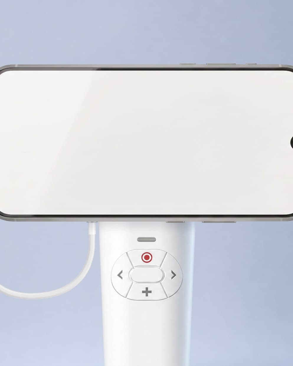
- Handheld, wireless, results on screen in seconds
- CE MDR class IIa medical device
Request a quote
- Training with every device
- Warranty & service
About
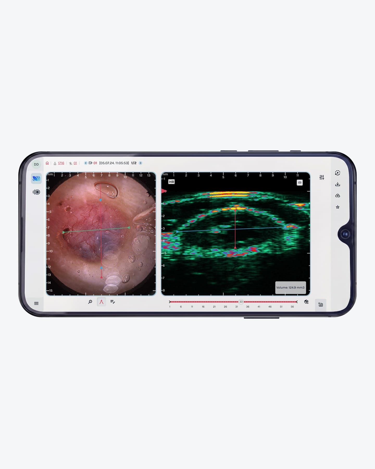
Dermus SkinScanner-M bridges the gap between surface dermoscopy and deep-tissue imaging. A real-time optical image guides the placement; a two-second scan then shows the full depth of the skin — epidermis, dermis, vessels, fillers and lesions — on the device and in the SkinAid platform. Objective information before, during and after treatment.
Treatment possibilities
Indications from the manufacturer's documentation. Each links to the treatment page.
How to use
Dermus skin scanner in the treatment room, step by step.
- 01
Position with optical guidance
Place the handheld on the skin. The built-in camera shows the area live at 10× magnification, so the scan line can be set precisely and repeated at follow-up.
- 02
Scan
Press once. The transducer sweeps mechanically across the field and captures the full depth of the skin in about two seconds.
- 03
Read the image on the spot
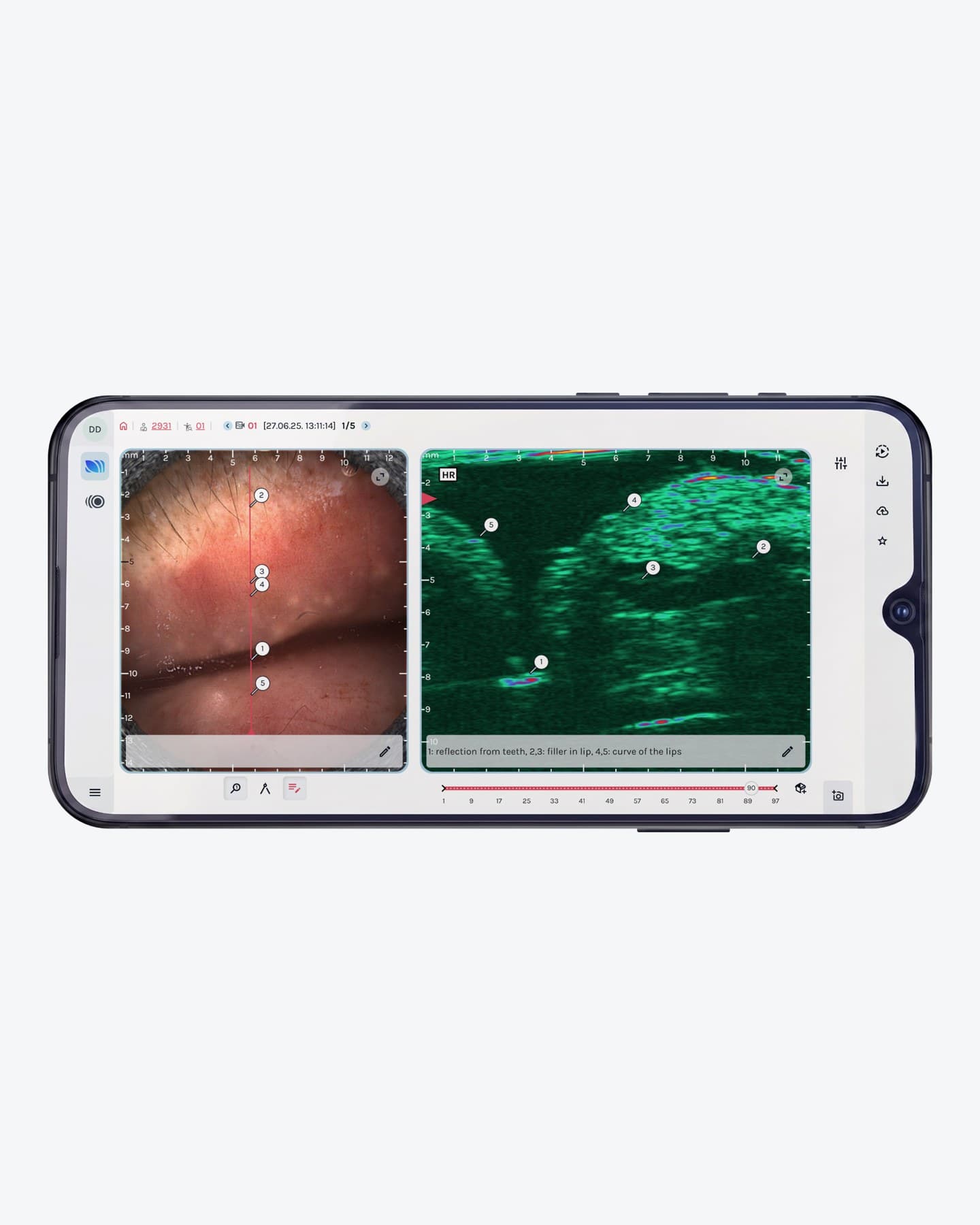
Optical and ultrasound images appear side by side on the screen: skin layers, thickness, lesion extent, vessels and injected material.
- 04
Analyse in SkinAid
Recordings are saved to the cloud. The web platform adds automated layer segmentation, correlated annotation, measurements in all dimensions and volume estimation.
- 05
Document and compare
Quantify skin changes over time and use the images in consultations, treatment planning and follow-up.
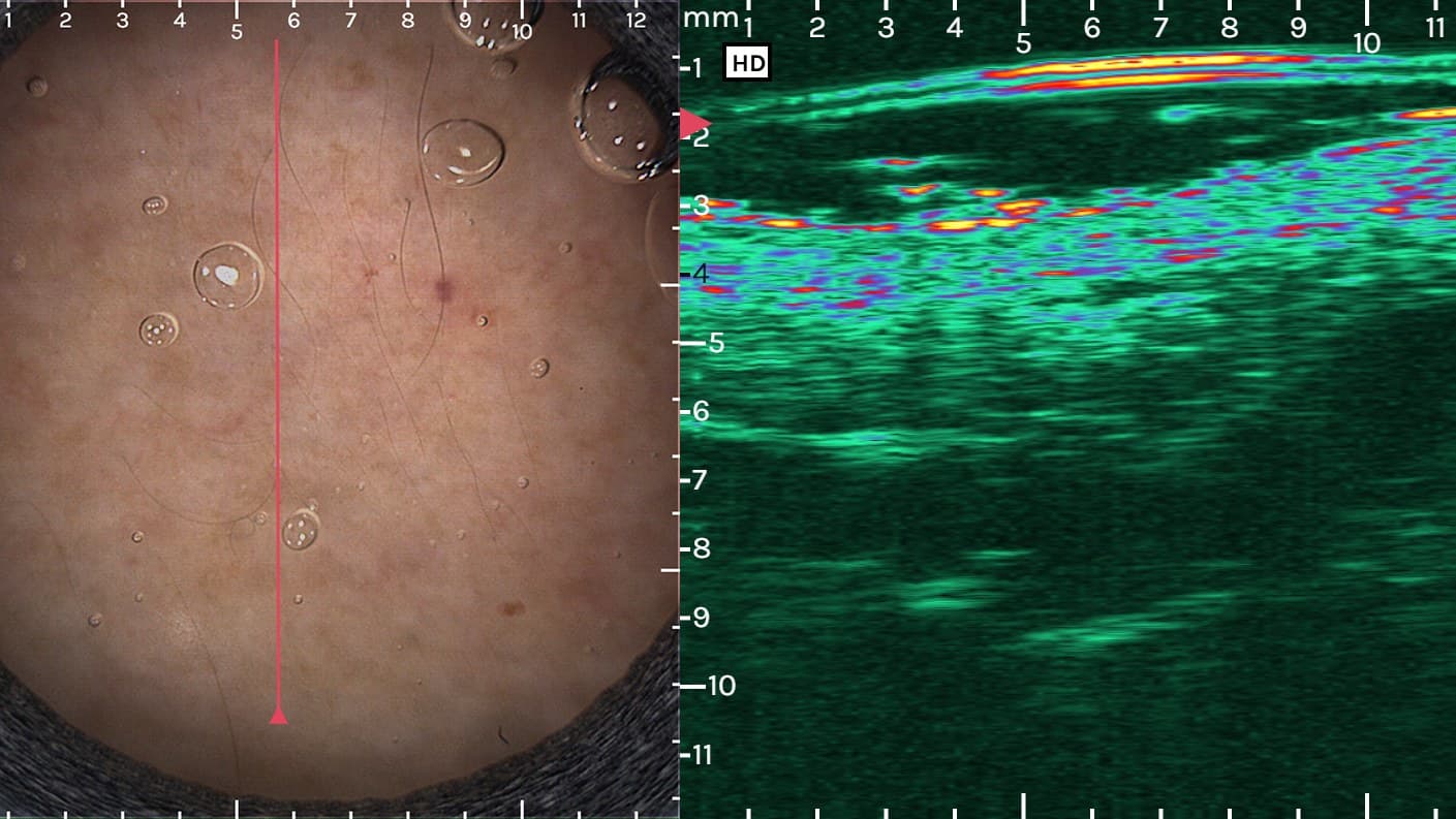
Dermal filler: dermoscopy and ultrasound show the filler deposit and its depth · Dermus
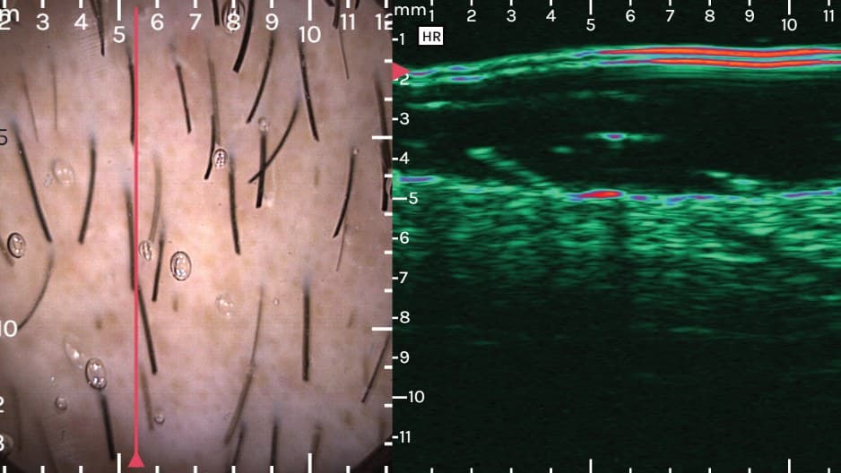
Hair and scalp: follicle depth and density · Dermus
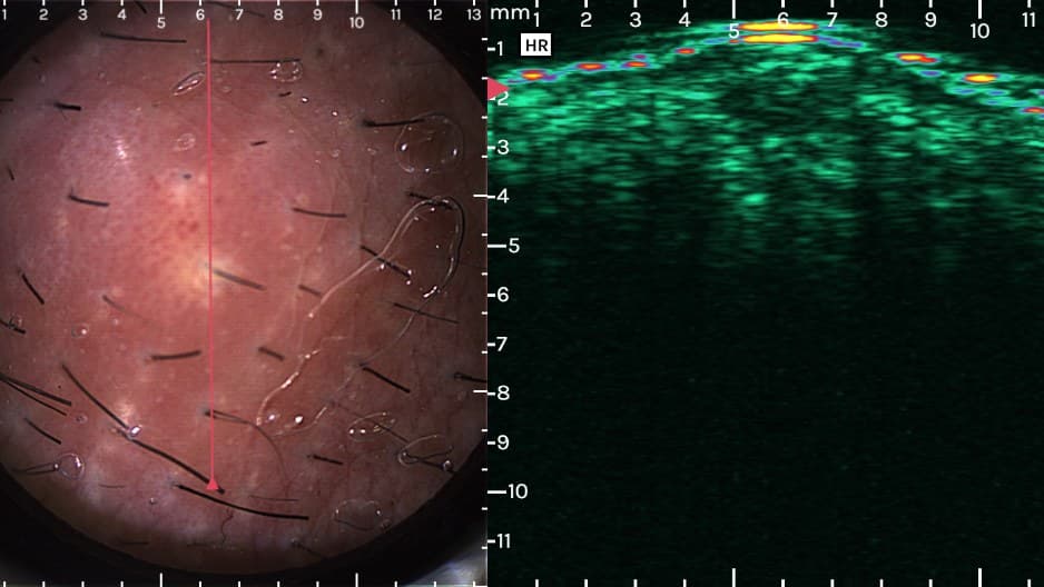
Acne: inflammatory lesions below the surface · Dermus
Detailed information
Technology, indications and specifications as documented by Dermus.
- Optically guided ultrasound
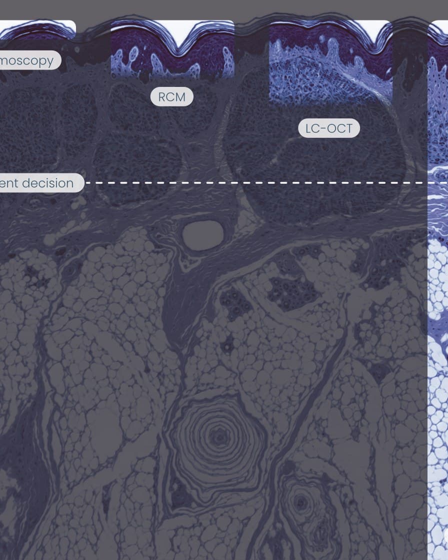
- The SkinScanner-M combines a dermoscopy camera with a 20–40 MHz single-element ultrasound transducer. The optical image guides positioning; the ultrasound shows the tissue beneath — down to 10 mm, well past the reach of dermoscopy, RCM or LC-OCT.
- In aesthetic practice
- Visualise hyaluronic acid filler and its position relative to vessels in lips and chin, monitor healing after CO₂ laser, assess androgenetic alopecia, acne, cysts and dermatofibromas, and image nail and scalp.
- In dermatology and oncology
- Used in hospitals and research centres for melanoma thickness estimation, basal cell carcinoma subtypes and margins, psoriasis, hidradenitis suppurativa and rosacea. Trusted by Erasmus Hospital, Roswell Park, Semmelweis University and Beiersdorf.
- SkinAid platform
- Web-based analysis with AI-assisted segmentation of skin layers, correlated annotation between optical and ultrasound images, lesion measurements, volume estimation and quantification of change over time.
- Made in Budapest
- Dermus Ltd. was founded in 2018 as a spin-off from Pázmány Péter Catholic University. ISO 13485 certified; SkinScanner-M received MDR certification in 2024 and is sold in more than 20 countries.
- Imaging
- Optically guided high-frequency ultrasound (dermoscopy + HFUS), 2-D power Doppler
- Ultrasound
- 20–40 MHz, 33 MHz nominal centre frequency, single-element transducer
- Depth
- Up to 10 mm
- Resolution
- 50–200 µm (frequency dependent)
- Field of view
- Ultrasound 12 × 12 mm · optical 15 × 15 mm at 10× magnification
- Acquisition
- Mechanical scan, about two seconds per recording
- Form factor
- Handheld, wireless, battery powered, 470 g, on-device display
- Data
- Automatic anonymised cloud storage · SkinAid web platform
- Regulatory
- CE, class IIa under Regulation (EU) 2017/745 · ISO 13485:2016
- Manufacturer
- Dermus Ltd., Budapest, Hungary
Publications
- Preliminary clinical experience with a novel optical–ultrasound imaging device on various skin lesions — Diagnostics, 2022doi:10.3390/diagnostics12010204
- Assessment of basal cell carcinoma using dermoscopy and high frequency ultrasound examination — Diagnostics, 2022doi:10.3390/diagnostics12030735
- Optically guided high-frequency ultrasound shows superior efficacy for preoperative estimation of Breslow thickness in comparison with multispectral imaging — Cancers, 2024doi:10.3390/cancers16010157
Talk to us